
")


 |
Axes and planes of a pennate diatom
|
Description of the trajectories
The use of video recordings for observing diatoms was already discussed in detail in Lesley A. Edgar (1979) and Ayumu Murasea et. al (2011). In both publications, diatoms are treated as point-shaped objects whose position can be described by the coordinates in the plane. In view of the sometimes complex movements of the diatoms, a complete kinematic description would have to capture the position of the diatoms in space. The basic axes and planes of a pennate diatom are schematically shown above (click to enlarge).
The ends of the diatoms (apices) lie at the points A and C. The valves were deliberately not represented as planes, in order to indicate that there is not necessarily only a single stable position in which the diatoms lie on a valve.
The next section is restricted to trajectories or sections of trajectories in which the diatoms do not erect, nor change between valve and girdle views. In the observation period, the valvar plane or the apical plane should lie in a good approximation parallel to the substrate.
Some diatoms (Navicula) are usually observed in valve view, some mostly in girdle view (Nitschia sigmoidea). Others often alternate between these positions, such as Cymatopleura solea. The ability to move can be significantly different in these two positions. Diatoms, which lie on a girdle face, can move freely provided their raphe is in mechanical contact with the substrate. If the raphe does not have this contact, as in the case of Pinnularia, only back and forth movements are possible in which a lump of extracellular polymeric substances (EPS) adheres to the substrate as well as to the raphe. The lump acts as an artificial substrate and couples the diatom to the substrate (MA Harper & JF Harper (1967)). In the 16x time-lapse video on the left you see a diatom of the genus Pinnularia (Length: approx. 220 µm) in valve view, which moves by means of two lumps of mucilage. These were made visible with Indian ink. The marking with ink also clearly shows the activity of the raphes. This old method will be discussed elsewhere on this site.
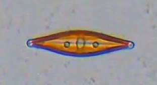
The requirement that the valvar plane or the apical plane is parallel to the plane of the substrate is rarely met perfectly. The small pictures of Craticula cuspidata show an example of deviations from this condition.
 |
 |
 |
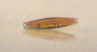
The first picture on the left shows the typical valve view, which is taken up from the vertical direction onto the substrate, i.e. perpendicular to the valvare plane. When Craticula cuspidata moves with a uniform motion, it is raising as is seen in the second picture when looking perpendicular to the apical plane. You see the diatom from almost horizontal direction as it is moving to the right, quasi "from the side". As its mirror image can be clearly seen on the substrate, the twice angle of inclination against the horizontal can be easily measured. In this case, the inclination is about 7.5°, but even 10° is not unusual. The observed length in valve view then appears foreshortened by the factor of the cosine of the inclination angle. At this small angle this results in less than 1% shortening, which is not relevant in the valve view and meets the given boundary condition sufficiently. There is a separate contribution about observation of Craticula cuspidata from a horizontal perspective.
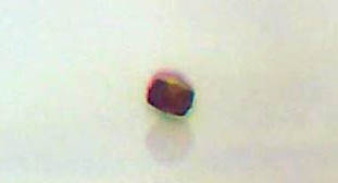
The third picture shows the diatom viewed perpendicularly to the transapical plane. It moves towards the observer. Here, in the video from which the picture was taken, different lateral inclination angles in sequence can be observed. If the diatom rotates 90 ° around the apical axis, it is lying on the girdle band. Such tilting may occur at the points of movement reversal during movement and as a result of collisions between diatoms. This is to be excluded within the analyzed section of a trajectory.
Formal description of the trajectory
For the mathematical description of the trajectory, the following coordinates are appropriate under the constraints described above (diatom lying on a valve with low inclination to the substrate):
- coordinates of the center of the diatom and the direction of the apical axis
- coordinates of the two apices
The choice of the cartesian coordinate system results in practice from the video recording.
Ayumu Murasea, Yosuke Kubotaa, Shigeyuki Hirayamaa, Yoshikazu Kumashirob, Teruo Okanob, Shigeki Mayamac, Kazuo Umemura (2011) Two-dimensional trajectory analysis of the diatom Navicula sp. using a micro chamber, Journal of Microbiological Methods, Volume 87, Issue 3, Pages 316–319
Lesley A. Edgar (1979) Diatom locomotion: Computer assisted analysis of cine film, British Phycological Journal, 14:1, 83-101, DOI: 10.1080/00071617900650111
M.A. Harper & J.F. Harper (1967) Measurements of diatom adhesion and their relationship with movement, British Phycological Bulletin, 3:2, 195-207, DOI: 10.1080/00071616700650051