
")


Observation with horizontal viewing direction
In connection with the detection of trajectories, an unusual camera angle on diatoms has already been mentioned, in which the viewing direction is inclined at a slight angle to the plane of the substrate. Here, one can recognize aspects in which the contact area between the diatom and the substrate is essential. The interaction between contact surface and trajectory has already been discussed.
After the explanation of the recording technique the following contributions will deal with the observation of certain genera and species. This method is also used elsewhere on this homepage, as in the contribution to the curvature of the trajectories of Surirella biseriata.
Vertical view
In the observation of living diatoms in the light microscope one sees them with a perpendicular view onto the substrate, ie in a vertical view. In the case of an upright microscope, you can either look at the top of the slide (bird's eye view) or with the inverse microscope from below into the culture vessel.
Horizontal view
If one looks at the diatoms from the perspective of a fictional tiny observer standing on the substrate, one sees them in a horizontal view. The word "side view" does not describe the situation quite correctly, as the diatoms can take different positions to the substrate. It is useful to look at the substrate with a slight angle, because then one can recognize the mirror image of the diatoms. This helps with measurements, because usually the horizon line is not clearly recognizable.
Benefits of the horizontal view
Probably the most important aspect is that the contact with the substrate becomes visible.
If you want to measure the inclination of the apical axis to the plane of the substrate, you could in a vertical viewing direction use the focus setting of the microscope at high magnification to determine the distance between the apices. Alternatively, the foreshortening of the distance between the apices can be measured. The first possibility is not suitable for video recordings, the second one is very imprecise at small inclinations as the shortening is proportional to the cosine of the inclination angle. From a horizontal view, the inclination of the apical axis is immediately recognizable.
Setup
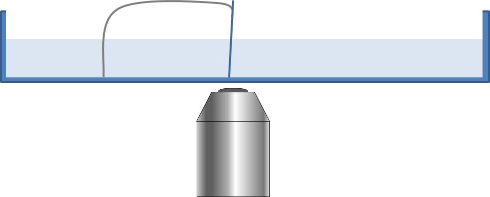
In a particularly simple method for recording with a horizontal view, a cover slip is placed almost vertically in a petri dish filled with water. A wire bow is attached to the cover glass and serves as a supporting leg. A sketch is shown on the left. If sufficient diatoms are brought into the petri-dish, then after a while diatoms will have migrated to the cover slip. If necessary, one can place the cover slip flat on the bottom of the petri dish and then carefully tilt it into the vertical position.
A more favorable setup for such observations would be an inverted microscope, which is tilted by about 90°, so that the optical path through the sample is almost horizontal. The observation takes place in a cuvette in which a transparent, very thin substrate (film) is arranged horizontally above the bottom so that the illumination beam path is only slightly influenced. The diatoms can then simply be put on the substrate and observed. So far I have avoided the effort to build this.
Difficulties
It is not surprising that the horizontal view is not widespread in light microscopy because one has to struggle with some difficulties:
- The distance between the bottom of the petri dish and the diatom can be very large. If this distance becomes too big, you can no longer focus the diatom. Even before reaching the maximum working distance, there is a water layer between the bottom of the petri dish and the diatoms, for which larger magnifying objectives are not corrected.
- The distance of the diatoms from the substrate can change considerably during the course of an observation. This requires frequent focusing.
- The illumination beam path is disturbed by the inclined cover slip.
An example
Diatoms of the species Cymatopleura solea (length approx. 80 µm) are considered as examples.
The video (60x time lapse) on the left shows a culture under the inverse microscope at different magnification levels. The optical axis is perpendicular to the substrate. It can be seen that the diatoms lie partly on the valves and partly on the girdle bands. There are changes between the positions, spontaneously or triggered by collisions.
A video recording (5x time lapse) from a horizontal perspective with respect to the substrate can be seen on the left below. The frame format has been changed by removing the top and bottom edges of the image. The inhomogeneous illumination therefore does not disturb the visual impression. The video shows a diatom which moves almost perpendicular to the viewer and changes its position to the substrate twice. Because the optical axis is almost perpendicular to the apical axis and the diatom has a small depth at this sight, the picture was taken successfully with a 10x objective. The focusing had to be adjusted only slightly, for example after the diatom tilted around its apical axis. The diatom moved at a short distance from the bottom of the petri dish.
Pictures from this perspective are informative when they show the Cymatopleura solea with a view parallel to the apical axis, i.e. perpendicular to the transapical plane. The third video on the left (20x time lapse) shows a diatom from this point of view (full screen mode is recommended). It shows that the diatom does not lie flat on its valve, but is tilted significantly around the apical axis. Therefore, the raphe can only be in contact with the substrate on one side of the apical axis. This inclination could be a consequence of the convex shape of the valve. A simple relationship between the direction of curvature of the trajectory and the inclination does not seem to exist in Cymatopleura solea.
Observation on natural substrate
The described method shows the movement of diatoms on a nearly vertical smooth glass surface. Observations in a natural environment tend to be made by chance.
In the 4x time lapse video on the left is a sample of a pond with a fiber on which a diatom moves. The contact between diatom and fiber is visible. Such images of natural samples can unfortunately hardly be used without an assignment to the species. In addition, the desired observation conditions are met only for a short time. In particular the direction from which the diatom is viewed changes frequently. Other objects can block the view onto the object of observation.