
")


Automixis in Cymbella aspera
In July 2017 I isolated diatoms of the genus Cymbella found in a pond in Hohenheim (Stuttgart, 48°42'34.0"N 9°12'29.1"E). The size of the diatoms at the time of isolation was about 240 µm and was in the range of 150 to
 |
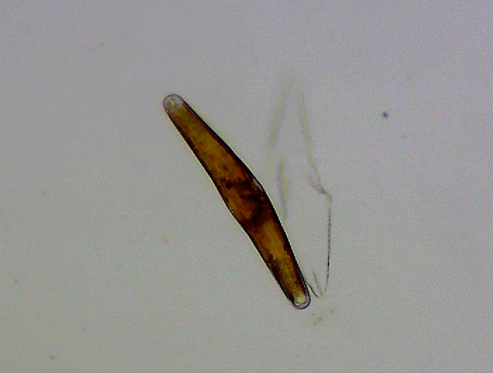
180 µm after 5 months in culture. The picture of a valve with a length of 151 µm can be seen in the upper half of the picture on the left. I assume it is Cymbella aspera.
At several locations in a culture dish, sexual reproduction began at this diatom length. Auxospores with attached valves and initial cells were found.
The size of the initial cells was not uniform. They had a length between 270 µm and 302 µm, whereby a length of 280 µm was very common. These initial cells were considerably longer than expected for this species. The ratio of length from the initial cell to a small cell showing sexual reproduction is
 |
approximately 1.8 to 2. In the picture on the top left below the small diatom a large diatom (length 270 μm) is shown, which originates from the first culture, which has developed from an initial cell. So it is at most a few generations away from the initial cell.
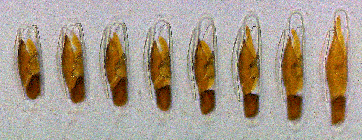
A typical image of a culture is shown in the image on the left (click to enlarge). Here you can see in several places evidence of sexual reproduction. A closer look reveals that each auxospore comes from only one cell. In a single diatom, two gametes are formed by meiosis, which then fuse to form a zygote (auxospore). It is a self-fertilization, or more precisely automixis. The valves of the diatoms open and an auxospore grows up. Automixis in Cymbella aspera was described by Lothar Geitler in "Automixis, Geschlechtsbestimmung und Pyknose von Gonenkernen bei Cymbella aspera" (Planta 1956, Vol. 47. pg. 359-373).
The auxospore is enclosed in a structured membrane, the so called perizonium. Inside the perizonium the auxospore grows and develops new frustules. The frustules of the original diatom stick to it from the outside. Finally, a finished diatom of maximum size leaves the perizonium. An almost finished initial cell can be seen subsequently on the left (click to enlarge). The cell has still not left the perizonium yet. In the picture to the right of it this already happened (click to enlarge).
 |
 |
The structure of the perizonium is clearly visible in phase contrast, but not in the brightfield. The following picture
 |
shows the brightfield and phase contrast alternately (2 seconds and 5 seconds).
In culture it turned out that the gametes often died after their formation and there was no fusion of the gametes. This happened far more often than the successful formation of an initial cell. Some of the initial cells also proved to be not viable or not cultivatable.
The continuous observation of the formation of an auxospore turned out to be difficult. In both Cymbella cistula and Cymbella aspera, it was striking that auxospores often died during several hours of observation. I suspect that auxospores have a very high sensitivity to high light intensities as they are typically used for microscopic observation. A video of a growing auxospore can be seen below:
The shown fragment is taken from a long-term observation and extends over a period of 84 hours. To protect the auxospore, the intensity of the light was kept extremely low, which leads to a strong image noise. In the beginning, the day-night rhythm was maintained as in cultivation. The nights are indicated by a short dark period in the video. After the second phase of darkness it became clear that the development had progressed in darkness. After that no dark phase was used. As the diatom and the auxospore do not lie parallel to the bottom of the petri dish, the lower part of the diatom is out of focus (objective with 20 x magnification). In the following picture, single images are placed next to each other with a time interval of about 4 hours (250 minutes):
 |
The superimposed lines show that the auxospore grows linearly over time within the limits of the image resolution. The growth rate was about 3.4 µm per hour.